Introduction: Lumbosacral plexopathy is a complex neurological condition involving dysfunction of the lumbosacral plexus, a network of nerves that innervates the lower extremities. It presents with symptoms such as pain, weakness, sensory disturbances, and impaired mobility, which can significantly impact an individual’s quality of life. Accurate diagnosis of lumbosacral plexopathy is essential for guiding appropriate treatment strategies and optimizing patient outcomes. Electromyography (EMG) and Nerve Conduction Studies (NCS) serve as invaluable tools in the diagnostic evaluation of this condition, aiding in localization of nerve involvement, differentiation from other neuropathies, and assessment of disease severity.
Understanding Lumbosacral Plexopathy: The lumbosacral plexus is formed by the ventral rami of the lumbar and sacral spinal nerves, giving rise to major nerves such as the femoral, obturator, sciatic, and pudendal nerves. Lumbosacral plexopathy encompasses a spectrum of disorders characterized by damage or dysfunction of these nerves, leading to motor, sensory, and autonomic deficits in the lower limbs. Etiologies of lumbosacral plexopathy include trauma, compression, inflammation, infection, radiation injury, neoplastic infiltration, and metabolic disorders.
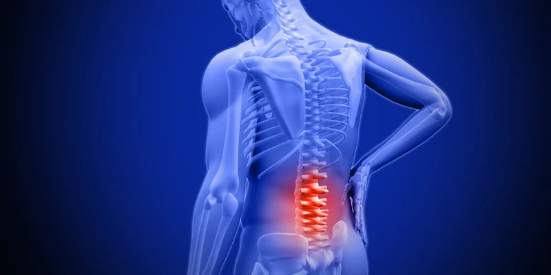
Clinical Presentation and Diagnostic Challenges: Patients with lumbosacral plexopathy typically present with symptoms localized to the lower back, buttocks, thighs, and legs. Common manifestations include pain radiating along the distribution of affected nerves, muscle weakness, sensory loss, altered reflexes, and impaired bladder or bowel function. However, the clinical presentation can vary widely depending on the underlying cause and the specific nerves involved. Differential diagnosis includes lumbar radiculopathy, peripheral neuropathies, spinal cord lesions, and musculoskeletal disorders, highlighting the importance of a thorough diagnostic workup.
Role of EMG/NCS in Diagnosis: EMG and NCS are essential neurophysiological tests utilized in the evaluation of lumbosacral plexopathy, providing valuable information about the integrity and function of peripheral nerves and muscles.
- Electromyography (EMG): EMG involves the insertion of small needle electrodes into targeted muscles to assess their electrical activity at rest and during voluntary contraction. In lumbosacral plexopathy, EMG findings may reveal denervation changes indicative of nerve injury or muscle dysfunction secondary to nerve compression or irritation. By analyzing the pattern, amplitude, and recruitment of motor unit potentials, EMG helps localize the site of nerve involvement and differentiate between primary neuropathic processes and secondary muscle pathology.
- Nerve Conduction Studies (NCS): NCS measures the speed and amplitude of electrical signals transmitted along peripheral nerves, assessing nerve conduction velocity, latency, and amplitude of sensory and motor responses. In lumbosacral plexopathy, NCS can identify abnormalities such as slowed conduction velocities, prolonged distal latencies, reduced sensory or motor amplitudes, and conduction blocks suggestive of nerve compression, demyelination, or axonal injury. By performing selective stimulation of individual nerves and recording responses from distal muscles or sensory nerves, NCS helps localize the site of nerve dysfunction within the lumbosacral plexus.
Localization of Nerve Involvement: EMG and NCS findings in lumbosacral plexopathy can provide valuable insights into the specific nerves affected and their anatomical distribution. Depending on the pattern of abnormalities detected, clinicians can infer the level and extent of nerve involvement within the lumbosacral plexus. For example, isolated involvement of the femoral nerve may suggest compression or injury at the level of the inguinal ligament or pelvic brim, while combined deficits affecting the sciatic and pudendal nerves may indicate more proximal pathology involving the sacral plexus or lumbosacral roots.
Challenges and Limitations: Despite their utility, EMG and NCS have certain limitations in the diagnosis of lumbosacral plexopathy. Variability in test interpretation, technical factors, and the presence of confounding factors such as pre-existing neuropathies or musculoskeletal disorders can impact the accuracy of results. Additionally, false negatives can occur, particularly in cases of early or mild nerve dysfunction where structural changes may be subtle. Therefore, clinical correlation and a comprehensive evaluation incorporating history, physical examination, and imaging studies are essential for accurate diagnosis and management.
Conclusion: Electromyography and Nerve Conduction Studies play a crucial role in the diagnostic evaluation of lumbosacral plexopathy, enabling clinicians to localize nerve involvement, differentiate from other neuropathies, and assess disease severity. By providing objective neurophysiological data, these tests complement clinical findings and guide appropriate treatment strategies tailored to the underlying etiology and extent of nerve injury.
Contact us today for more information.
Photo by: David Cross